Implantes Dentales en Panamá
Dra. Mariulys Ramos, Especialista en Periodoncia e Implantes Dentales
La marca de implantes MIS es una de las más reconocidas en el mundo por poseer altos estándares de calidad en todas las líneas y marcas de implantes.
Dra. Mariulys Ramos, Especialista en Periodoncia e Implantes Dentales
La marca de implantes MIS es una de las más reconocidas en el mundo por poseer altos estándares de calidad en todas las líneas y marcas de implantes.
La base del éxito, consiste en hacer desde un principio una evaluación correcta del hueso remanente para saber si se puede poner un implante o si requiere tratamiento previo para que el implante se osteointegre adecuadamente.
Nacida en Panamá, Especialista en Periodoncia e Implantes, Graduada de la Prestigiosa Universidad de Buenos Aires. Cirujano Dental graduada en la reconocida Universidad de Panamá.
Se ha destacado tanto nacional como internacionalmente, en su práctica privada, la docencia y diversas actividades sociales relacionadas con su vocación.
Como presidenta de la sociedad está encargada de organizar la XV Jornada Internacional de Periodoncia, el cual posiciona a Panamá como un referente de la región.
Más de 6 años como docente en la Universidad Latina de Panamá.
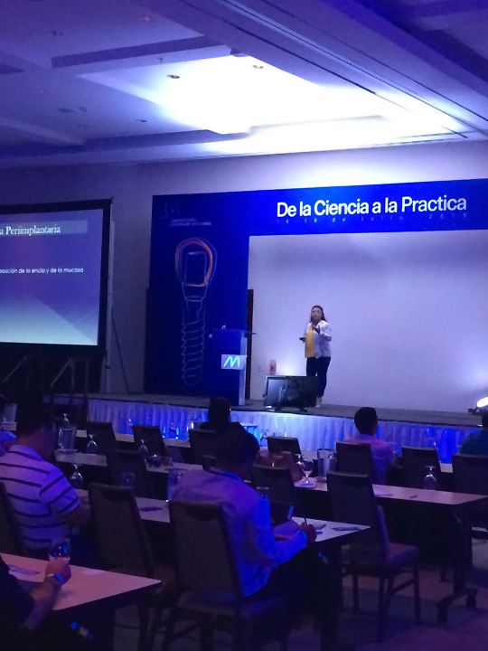
En la foto se la vé exponiendo en el I Simposio MIS Centro América y Caribe de la Ciencia a la Práctica - Hotel Westing Golf, Costa Rica.
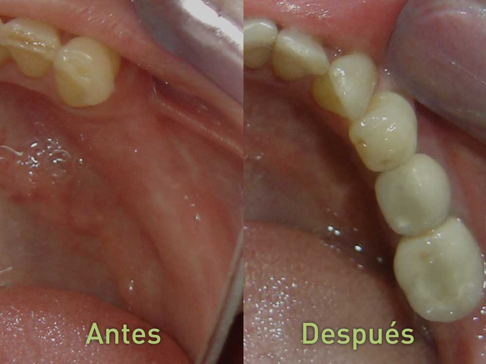
El implante se une al hueso y no a otros dientes. Ejerciendo una fuerza similar a los dientes naturales permitiendo mantener su forma biológica.
En ningún momento del procedimiento se siente dolor ni molestias. El postoperatorio suele ser menos traumático que otros procedimientos.
La vida útil de un implante generalmente es de al menos 20 años, pudiendo en la mayoría de los casos tener una duración mayor.
El implante dental mantiene la funcionalidad original del diente, evitando la pérdida ósea que ocurre con las alternativas más comunes.
La osteointegración ocurre a nivel directo molecular de la superficie de titanio del implante con el hueso, en un período de 6 a 8 semanas.
La sensación masticatoria es similar a la de un diente natural,y en casos de pérdida completa son una excelente alternativa a una dentadura postiza.
Su mandíbula comienza a achicarse por la falta de estimulación. La pérdida de los dientes afectará su sonrisa y cambiará la forma de su rostro, lo que hace que se vea prematuramente envejecido.
Contáctenos hoy mismo y solicite una cita.